Nightly Pain in the Thigh – Think of Osteoid Osteoma
Nightly Thigh Pain
A 5 year old girl presented to me with nightly pain in her left thigh for the last 3 months. Her mother gave the story that her daughter would wake up at about 3am each night with severe pain in her left thigh. She would cry and would not be able to stand or walk on that leg. This pain was relieved by taking syrup Brufen (Ibuprofen).
They had previously consulted other doctors who did blood tests and x-rays of her left thigh and was told that everything was normal.
Her pain was attributed to “growing pains” by her doctors.
There was no abnormality of her left hip or left thigh when I examined her in my office. She was able to walk, run and jump without pain.
I ordered an x-ray of her pelvis and left thigh (femur bone).
Subtle Features on X-rays
The x-rays of her pelvis allowed me to compare the hip joints of both the right and left sides.
It clearly showed increased sclerosis (thicker bone) in the left hip around the lesser trochanter.

The arrow shows the area of increased whiteness or sclerosis of the bone from a possible osteoid osteoma.
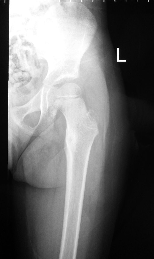
The x-rays were suggestive of a condition called Osteoid Osteoma.
A CT-scan was immediately ordered to confirm the diagnosis.
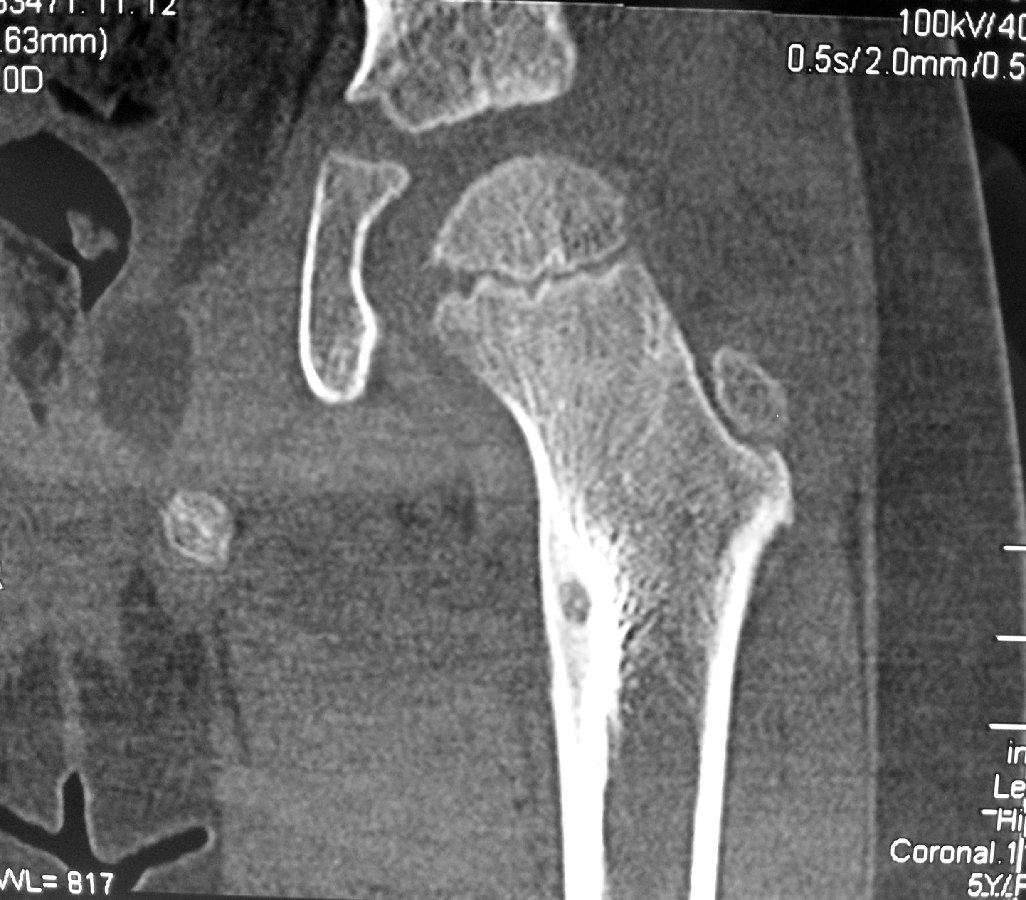
CT-scan shows a nidus of Osteoid Osteoma in the Lesser Trochanter of the Left Hip Joint

What is Osteoid Osteoma?
- An osteoid osteoma is a benign bone tumor that arises from osteoblasts.
- Osteoid osteomas tend to be less than 1.5 cm in size.
- The tumor can be in any bone in the body but are most common in long bones, such as the femur and tibia.
- They account for 10 to 12 percent of all benign bone tumors.
- “Osteoid osteomas may occur at any age, and are most common in patients between the ages of 4 and 25 years old.
- Males are affected approximately three times more commonly than females
What Are the Symptoms?
The most common symptom of an Osteoid Osteoma is dull pain that escalates to severe at night.
Other symptoms include:
- limping
- muscle atrophy
- bowing deformity
- swelling
- increased or decreased bone growth
Treatment
Pain may be relieved by aspirin or other. nonsteroidal antiinflammatory drugs e.g. Brufen.
Percutaneous radiofrequency ablation is the preferred treatment option.
This is a minimally invasive procedure in which radio frequencies are passed beneath the skin through a needle to kill the tumor cells by heating them to a high temperature.
This technique is performed under general anesthesia and CT-fluroscopic guidance and does not weaken the bone as much as surgery does.
The recovery time is also shorter for this treatment.
Percutaneous Radiofrequency Ablation of Osteoid Osteoma
This girl was crying in pain by the time the CT-scans were done. It was obvious that something was wrong. The CT-scan confirmed the presence of an osteoid osteoma of the lesser trochanter of her left hip with pain over her left thigh.
She underwent percutaneous radiofrequency ablation of the tumour under general anaesthesia.
This procedure is done under CT-fluoroscopic guidance.
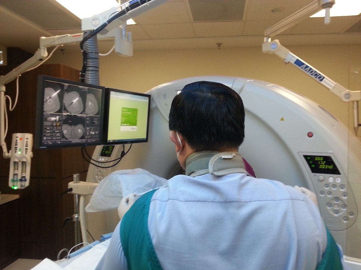
Dr HC Chang using CT-fluroscopic guidance
A 16F needle is inserted through the skin and advanced in a safe zone into the bone of the lesser trochanter under CT-guidance.

The radiofrequency probe is then inserted into the bony tumour.
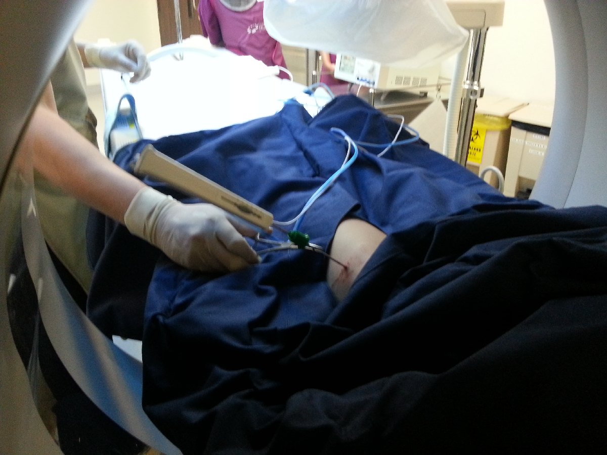
Correct placement of the radiofrequency probe into the bony tumour lesion is confirmed on CT-scan.

Heat energy is generated by this radiofrequency machine. About 6 minutes worth of energy at 90 degreec Celcius is used to burn the lesion.

Recovery
The patient is able to walk the following day with minimal discomfort. The pain in her left thigh from the tumour goes away almost immediately after the procedure.
The success rate of this procedure is in the region of 80%. In some patients, it may need to be repeated a 2nd time. The success rate improves to over 90% with the 2nd treatment.
For more information on osteoid osteoma or treatment of this tumour, please contact us at +65-683 666 36 or visit our website at http://www.ortho.com.sg
